[HIDE]
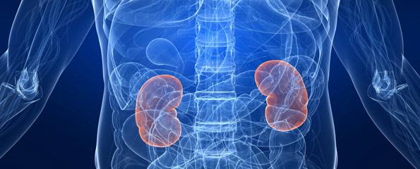
Renal
cell carcinoma is the most common cancer of the kidney, comprising about 75 per
cent of the renal cancers. It is also known as hypernephroma and Grawitz
tumour. Renal cell carcinoma usually occurs during the age of 50 to 60 years.
The incidence is twofold higher in males as compared to females. It mostly
affects the people living in urban and industrialised areas. Renal cell
carcinoma is an adenocarcinoma, usually arising from the cells of proximal
tubules in the renal cortex. The exact cause of renal cell carcinoma is not
fully understood. Researchers believe that smoking is linked to about 30 per
cent cases of the renal cell carcinoma. It has been observed that the workers
of rubber, leather, petroleum, textile, dye, insulation and plastic industries
are more likely to develop renal cell carcinoma. Risk factors include obesity
and exposure to radiation & chemical carcinogens that might have been
inhaled, ingested or produced within the body. Prolonged abuse of analgesics
may lead to renal cell carcinoma.
Patients
suffering from renal cell carcinoma usually, remain asymptomatic in the early stages
of the disease. In advanced stages of the renal cell carcinoma, there may be
haematuria (gross or microscopic), pain in the flanks and palpable renal mass.
These three signs are collectively known as a classical triad. Other symptoms of
renal cell carcinoma include fever (without infection), nausea, vomiting, loss
of appetite, loss of weight, malaise, lassitude and anaemia. There may be
endocrine disturbances, polycythaemia, hypertension, nephrotic syndrome and
hypercalcaemia. Varicocele may occur due to occlusion of the renal and the
testicular veins. There may be occlusion of the inferior vena cava leading to
oedema in the legs. Renal cell carcinoma usually metastasises to the lungs,
bones and the para-aortic lymph nodes.
Staging
of the renal cell carcinoma is done as follows:
Procedures
used in diagnosis and evaluation of the renal cell carcinoma include urine
analysis, intravenous pyelography (IVP), intravenous urogram (IVU), renal
arteriography, ultrasound, CT scan, MRI and biopsy.
Nephroblastoma
(Wilm's tumour) is the second most common cancer of the kidney. It is a fast-growing tumour that usually affects young children during the first decade,
especially in the first year of life. Nephroblastoma comprises about 8 per cent
of the childhood tumours. It affects boys and girls equally. The exact cause of
nephroblastoma is not fully understood. It has been observed that about 10 per
cent cases of the nephroblastoma have some kind of congenital abnormality.
Researchers believe that there are hereditary and non-hereditary types of
nephroblastoma. The hereditary nephroblastoma occurs in younger children and
usually affects both the kidneys. The most common symptom of nephroblastoma is
a mass in the abdomen. Other symptoms include low-grade fever, loss of
appetite, loss of weight, pallor, lethargy and haematuria. Nephroblastoma
usually metastasises to the liver, lungs and the bones.
Staging
of the nephroblastoma is done as follows:
Procedures
used in the diagnosis and evaluation of nephroblastoma include urine analysis,
IVP, ureteropyeloscopy, selective renal arteriography, ultrasound, CT scan, MRI
and biopsy.

Disclaimer:
This content is for information and educational purposes only and should not be perceived as medical advice. Please consult a certified medical or healthcare professional before making any decision regarding your health using the content above.
Click here to go back to the list of all Articles


Kidney Cancer (Renal Cell Carcinoma and Nephroblastoma)